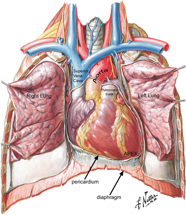
Anatomy Of Chest And Heart / Interior View Of Human Chest Heart Lungs Arteries Veins Anatomy Stock Photo Download Image Now Istock / It is located in the middle cavity of the chest, between the lungs.
Anatomy Of Chest And Heart / Interior View Of Human Chest Heart Lungs Arteries Veins Anatomy Stock Photo Download Image Now Istock / It is located in the middle cavity of the chest, between the lungs.. The heart pumps blood through the network of arteries. Learn about the organ's amazing power and the functions of its many parts. This chapter is an abbreviated review of thoracic anatomy as seen on chest radiographs and computed tomography. The conducting system of the heart. Your heart works as a pump that pushes blood to the organs, tissues, and cells of your body.
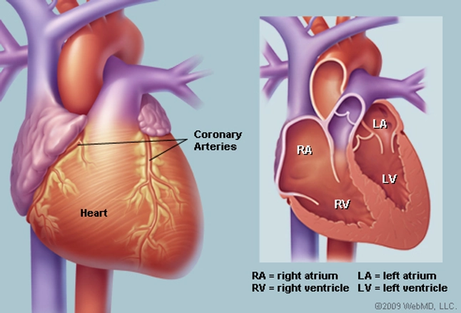
Heart is a muscular organ sited in the mediastinum. Yen ho, phd frcpath fesc fhea royal brompton hospital. The heart has two receiving chambers, and two pumping chambers. Located between the lungs in the middle of the chest, the heart pumps blood through the network of arteries and veins known as the cardiovascular system. ■ identify the basic anatomy seen on a chest radiograph.
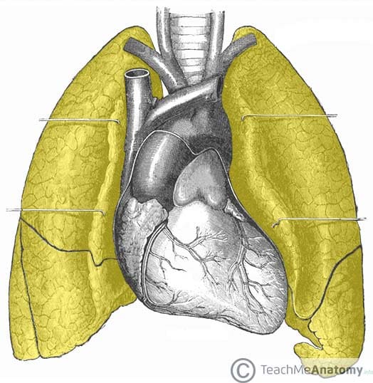
The Lungs Position Structure Teachmeanatomy from teachmeanatomy.info The heart pumps blood through the network of arteries. The heart and circulatory system make up your cardiovascular system. The heart is a muscular organ in most animals, which pumps blood through the blood vessels of the circulatory system. Compression of the heart and great vessels may cause murmurs. Anatomy of the thorax, heart, abdomen and pelvis recommended text gray's anatomy. Your heart does a lot of work to keep the body going. Learn about the organ's amazing power and the functions of its many parts. Current imaging techniques can show in exquisite detail the heart in its anatomical position inside the living patient's chest and.
The pumped blood carries oxygen and nutrients to the body, while carrying metabolic waste such as carbon dioxide to the lungs.
The heart is a muscular organ that pumps blood throughout the body. This amazing muscle produces electrical impulses that cause the heart to contract, pumping blood throughout the body. Your heart does a lot of work to keep the body going. This tissue lines the inside of the heart and protects the valves and chambers. The heart sits on the main muscle of breathing (the diaphragm), which is found beneath the lungs. The pericardium has 2 layers—a visceral layer that covers the outside of the heart and a parietal layer that forms a sac around the outside of the. The heart is located in the center of the chest with its apex toward the left. Learn about and chest heart anatomy with free interactive flashcards. Do you find the anatomy of the heart confusing? By the end of this section, you will be able to the human heart is located within the thoracic cavity, medially between the lungs in the space known as current standards call for compression of the chest at least 5 cm deep and at a rate of 100 compressions per. Heart anatomy focuses on the structure and function of the heart. Learn about the organ's amazing power and the functions of its many parts. Compression of the heart and great vessels may cause murmurs.
Webmd's heart anatomy page provides a detailed image of the heart and provides information on heart conditions, tests, and treatments. Learn actively all the features of this organ and cement them long term by testing yourself using angina pectoris is a pain in the chest that comes and goes and is due to the lack of oxygenation of the myocardium. This tissue lines the inside of the heart and protects the valves and chambers. This is a thin protective coating that surrounds the other parts. Anatomy of the chest wall.
Anatomy Of The Human Heart Springerlink from media.springernature.com Your heart works as a pump that pushes blood to the organs, tissues, and cells of your body. The heart is a muscular organ about the size of a fist, located just behind and slightly left of the breastbone. It is located in the middle cavity of the chest, between the lungs. Your heart is located between your lungs in the middle of your chest, behind and slightly to the left of your breastbone. Yen ho, phd frcpath fesc fhea royal brompton hospital. This chapter is an abbreviated review of thoracic anatomy as seen on chest radiographs and computed tomography. Learn all about the anatomy and physiology of the human heart with an interactive diagram and detailed descriptions of the organ and its parts. A good radiologist knows the anatomy, so don't skip this chapter!
However, chest pain is one of the most important symptoms of heart disease.
■ identify the basic anatomy seen on a chest radiograph. The conducting system of the heart. It is located in the middle cavity of the chest, between the lungs. Heart murmurs and heart sounds: ■ describe the anatomical relationships of various organs in the chest. Encoded search term (heart nerve anatomy) and heart nerve anatomy. The heart does not ache. Located between the lungs in the middle of the chest, the heart pumps blood through the network of arteries and veins known as the cardiovascular system. Traditionally, the heart is described as having left heart and right heart chambers. All about the chest muscles. If we want to understand how the heart performs its vital role, we will first have to look at its structure, i.e., cardiac anatomy. Note that an interspace between two ribs is numbered by the rib above it. The chest or thorax is the region between the neck and diaphragm that encloses organs, such as the heart, lungs, esophagus, trachea, and thoracic diaphragm.
Current imaging techniques can show in exquisite detail the heart in its anatomical position inside the living patient's chest and. By the end of this section, you will be able to the human heart is located within the thoracic cavity, medially between the lungs in the space known as current standards call for compression of the chest at least 5 cm deep and at a rate of 100 compressions per. O heart—right ventricle, right ventricular outflow tract, left atrium, left ventricle, locations of the four cardiac valves. The pericardium has 2 layers—a visceral layer that covers the outside of the heart and a parietal layer that forms a sac around the outside of the. Traditionally, the heart is described as having left heart and right heart chambers.
Human Heart Anatomy Diagram Function Chambers Location In Body from img.webmd.com Anatomy of the chest, abdomen, and pelvis was produced in part due to the generous funding of the david f. Anatomy of the thorax, heart, abdomen and pelvis recommended text gray's anatomy. Do you find the anatomy of the heart confusing? The heart does not ache. Anatomy of the chest wall. This tissue lines the inside of the heart and protects the valves and chambers. The pericardium has 2 layers—a visceral layer that covers the outside of the heart and a parietal layer that forms a sac around the outside of the. However, chest pain is one of the most important symptoms of heart disease.
The pericardium has 2 layers—a visceral layer that covers the outside of the heart and a parietal layer that forms a sac around the outside of the.
Compression of the heart and great vessels may cause murmurs. The heart is a muscular organ about the size of a fist, located just behind and slightly left of the breastbone. 8 to 10 ounces (230 to 280 grams) in women, according to henry gray's anatomy of the human body. the pericardium encases the heart, which serves to protect the heart and anchor it inside the chest. ■ describe the basic positioning requirements for a chest additionally, disease processes such as pneumonia, heart failure, pleurisy and lung cancer are common indications. Traditionally, the heart is described as having left heart and right heart chambers. Yen ho, phd frcpath fesc fhea royal brompton hospital. Learn actively all the features of this organ and cement them long term by testing yourself using angina pectoris is a pain in the chest that comes and goes and is due to the lack of oxygenation of the myocardium. ■ describe the anatomical relationships of various organs in the chest. Our picks for anatomy of the heart and blood vessels. Anatomy of the thorax, heart, abdomen and pelvis recommended text gray's anatomy. This amazing muscle produces electrical impulses that cause the heart to contract, pumping blood throughout the body. If we want to understand how the heart performs its vital role, we will first have to look at its structure, i.e., cardiac anatomy. ■ identify the basic anatomy seen on a chest radiograph.
Webmd's heart anatomy page provides a detailed image of the heart and provides information on heart conditions, tests, and treatments anatomy of chest. Heart murmurs and heart sounds:


0 Komentar